Medical Device Manufacturer Catalog No.8122
OROSOL® can produce medical device – Ophthalmic Imaging & Dry Eye Management Solutions.
Item Name: OROSOL® Ophthalmic Imaging & Dry Eye Management Solutions.
Brand: OROSOL®
OROSOL® can produce all kinds of medical devices.
If you are interested in this product, please feel free to contact us: RoboticWorkCell@outlook.com
Product Description
Medical Device Manufacturer Catalog No.8122
General View of OROSOL® Manufacturer No.8122:
OROSOL Machinery and Equipment Manufacturing LLP is located in China. With Manufacturer No.8122 founded in 2015 by a team of overseas PhDs, the company is a National High-Tech Enterprise and a leading specialist in high-precision ophthalmic optical devices. The company holds 94 valid patents, has obtained CE, FDA, and NMPA certifications. The company has completed nearly 100 million yuan in Series C financing and has 8 products approved for the Chinese market. Products are exported to multiple countries and regions worldwide.
Medical Device Manufacturer Catalog No.8122
| Product Name & Model | Parameters & Specifications | Features & Applications |
|---|---|---|
| OS-OCT Luminor-A80 Angiography | 1. Full-process intelligent diagnostic OCT, angiography version, for outpatient rapid screening of fundus disease. 2. Axial resolution 5 micron or better in tissue; horizontal resolution 13 micron or better. 3. Maximum A-scan speed 80 kHz with tolerance 5 percent; maximum scanning depth 3.5 mm in tissue with tolerance 3 percent. 4. Refraction compensation range -20 D to +20 D. 5. Light source centre wavelength 840 nm with tolerance 10 nm, half-peak width 35 nm with tolerance 5 nm, optical power 750 microwatt or less at the cornea. 6. Scanning range 13 x 13 mm in tissue with tolerance 5 percent; OCT field of view 42 by 42 degrees with tolerance 5 percent. 7. Fundus image light source centre wavelength 760 nm with tolerance 5 nm, optical power 50 microwatt or less. 8. Retinal thickness measurement tolerance 3 percent and repeatability 2 percent or better. 9. Angio OCT non-invasive flow imaging with intelligent retinal layer segmentation; voice navigation with one-key automatic capture and eye tracking. |
1. Used for tomography and examination of retinal, macular and optic disc structure. 2. Auxiliary diagnosis for diabetic retinopathy, age-related macular degeneration, central and branch retinal vein occlusion, choroidal neovascularization, polypoidal choroidal vasculopathy, central serous chorioretinopathy, macular hole and glaucoma. 3. Angiography mode shows choroidal and retinal microvessels layer by layer without contrast injection. 4. Suited to high-volume outpatient screening where examination time per patient is short. |
| OS-OCT Luminor-A60 Angiography | 1. Full-process intelligent diagnostic OCT, blood flow version, with voice-guided workflow. 2. One-key automatic capture; automatic alignment reduces operator dependence. 3. High-efficiency eye tracking keeps the scan centred during acquisition. 4. Intelligent segmentation algorithm separates retinal layers automatically for thickness analysis. 5. Angio OCT technology displays choroidal and retinal vessels in layers without contrast agent. 6. Single-line scan up to 13 mm centred on the macula images macula and optic disc in one pass. |
1. Used for detecting fundus neovascularization and early retinal lesions. 2. Layer-by-layer vessel display supports precise localisation of microvascular abnormality. 3. Thirteen millimetre line scan captures macula and disc together, reducing repeat captures in wide-field assessment. |
| OS-OCT Luminor-D60 Standard | 1. Full-process intelligent diagnostic OCT, standard structural version, with combined automatic and manual operation. 2. Lateral resolution 13 micron; scanning depth 3.5 mm reaching the choroid in highly myopic eyes. 3. Scanning range 13 mm covering macular and optic disc regions in one image. 4. High-speed OCT acquisition with layer thickness measurement on the dedicated analysis workstation. 5. Macular analysis with multiple scan patterns, binocular comparison and follow-up comparison. 6. Glaucoma analysis with ganglion cell complex thickness and retinal nerve fibre layer thickness. 7. Anterior segment diagnosis with visible corneal structure, manual or automatic corneal layer thickness measurement and angle scan alignment. 8. Cloud reading service providing remote interpretation of OCT images by reviewing specialists. |
1. Used as the routine structural OCT workhorse for retina clinics and screening centres. 2. Anterior segment mode extends use to cornea and angle assessment without a second device. 3. Remote reading lets sites without a resident retina specialist report images quickly. |
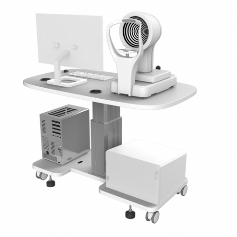
| OS-OCT OCTA2020 Table Model | 1. A medical device registration is held for this product. 2. OCTA2020 configuration comprises optical imaging unit, electronic control unit, positioning unit, computer, monitor, work table and software version V1.0. 3. Overall dimensions 850 x 1100 x 1000 mm including the table. 4. Phase variance algorithm provides blood flow imaging without contrast agent. 5. Layered display of microvessels at different fundus depths. 6. Single-line scan 11 mm wide centred on the macula, imaging macula and optic disc together. |
1. Used for tomography and examination of fundus tissue structure. 2. Non-invasive, non-contact high-resolution imaging of retina, macula and optic disc. 3. Auxiliary diagnosis for diabetic retinopathy, age-related macular degeneration, retinal vein occlusion, choroidal neovascularization and glaucoma. |
| OS-OCT OCTA2010 Compact Model | 1. Compact angiography OCT configuration comprising optical imaging unit, electronic control unit, positioning unit, computer, monitor and software version V1.0, without the work table. 2. Same optical engine and phase variance angiography algorithm as the OCTA2020. 3. Smaller installation footprint for rooms where a full table is not required. 4. Layered choroidal and retinal vessel imaging without dye injection. 5. Export of images and measurements to the reading workstation. |
1. Used for fundus structural and vascular examination in space-limited clinics. 2. Shares consumable-free operation with the table model, so no per-patient cost. 3. Suited to satellite clinics of a hospital network using the same reporting software. |
| OS-Vein Finder ZD-JM-260 Series | 1. Medical vascular imaging device using infrared detection of superficial subcutaneous veins with real-time in-situ projection of the vein image onto the skin. 2. Three configurations: hand-held ZD-JM-260-04, desktop ZD-JM-260-03 with hover bracket and multi-joint arm, and hand-push trolley stand. 3. Working principle: venous blood absorbs the infrared peak, a micro camera collects the image, photoelectric conversion produces the vein map and a micro projector returns it to the skin surface in situ. 4. Three image colour modes, blue and white, red and green, red and white, switched with one key. 5. Adjustable image brightness for different ambient lighting; unique image enhancement system for high accuracy. 6. Medical cold light source, no ionising radiation and no eye injury; touch sleep for fast power saving. 7. Hand-held version is compact and light and can also be fixed on the trolley bracket or a desktop bracket. |
1. Used for venipuncture in wards, outpatient departments, laboratories and emergency and first aid settings. 2. Most effective in children, obese patients, oedematous limbs and low blood volume patients with difficult vascular localisation. 3. Raises first-attempt puncture success, reduces patient pain and lowers phlebotomy-related complaints. 4. No X-ray or gamma radiation, so staff can use it at the bedside without shielding. |
| OS-Platform ZD Medical | 1. Established 19 January 2015; national high-tech enterprise focused on high-end ophthalmic equipment. 2. Product families: Luminor OCT and angiography OCT series, OCTA2010 and OCTA2020, and the ZD-JM-260 vascular imaging series. 3. Phase variance optical coherence tomography developed in house, able to detect diabetic macular change and retinal microvascular proliferation simultaneously; proprietary technology applied. 4. A medical device registration is held for this product. 5. Cloud reading platform and AI-assisted analysis offered alongside the imaging hardware. |
1. Supplies eye hospitals, ophthalmology departments and primary screening sites with OCT and vein visualisation equipment. 2. Hardware, analysis software and remote reading are supplied as one diagnostic chain. 3. Focused portfolio allows one service organisation to support both the imaging and the vein access products. |
Certificates of OROSOL® Medical Device Manufacturer 8122:
- ISO 13485:2016 — Medical Devices Quality Management System
- NMPA Medical Device Production License
- NMPA Medical Device Registration Certificate (Class II) — 8 products approved
- CE Marking — Compliance with European Medical Device Regulation
- National High-Tech Enterprise Certification (China)
- 94 Authorized Patents — 90+ effective patents
- RoHS Compliant — Restriction of Hazardous Substances Directive (EU)
- REACH Compliant — Registration, Evaluation, Authorization and Restriction of Chemicals (EU)
E-mail us to get a free quote: RoboticWorkCell@outlook.com

Address:
International Sales Department
OROSOL® Machinery and Equipment Manufacturing LLP
Address: Room A-522, No. 188 Yesheng Road, Lingang New Area, China (Shanghai) Pilot Free Trade Zone.
Tel: (+86) 173-2682-9204
E-mail: RoboticWorkCell@outlook.com


