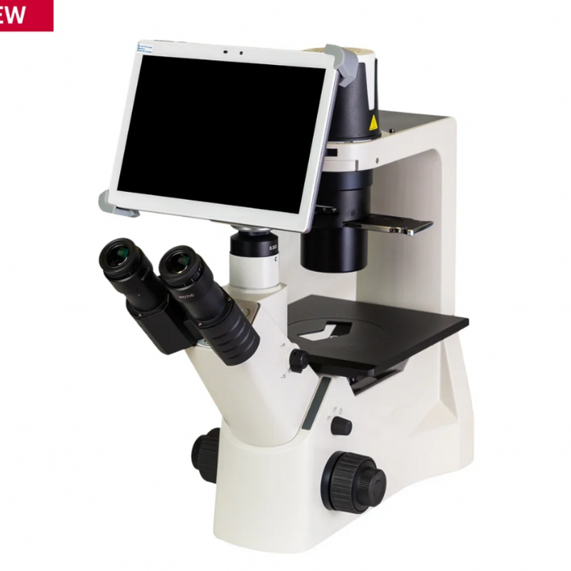
Medical Device Manufacturer Catalog No.8007
OROSOL® can produce medical device – Microscopy & Digital Pathology Solutions.
Item Name: OROSOL® Microscopy & Digital Pathology Solutions.
Brand: OROSOL®
OROSOL® can produce all kinds of medical devices.
If you are interested in this product, please feel free to contact us: RoboticWorkCell@outlook.com
Product Description
Medical Device Manufacturer Catalog No.8007
General View of OROSOL® Manufacturer No.8007:
OROSOL Machinery and Equipment Manufacturing LLP is located in China, with Manufacturer No.8007 a modern manufacturing facility spanning over 80,000 square meters. The company employs more than 1,200 professional staff members, including a dedicated R&D team of over 200 engineers and technicians specializing in optical microscopy, digital imaging, and digital pathology solutions. The company is a 100% wholly-owned subsidiary of a publicly listed company (Stock Code: 300341) and is one of the world’s leading manufacturers of microscopes and digital pathology systems. Products are exported to more than 70 countries and regions worldwide, serving millions of laboratories in education, research, healthcare, and industrial sectors. The company has obtained over 100 patent authorizations in optical, mechanical, and software technologies.
Medical Device Product Catalog No.8007:
| Product Category | Product Name & Model | Parameters & Specifications | Features & Applications |
|---|---|---|---|
| Digital Pathology Scanners | OS-EDS-1 EasyScan 1 Digital Slide Scanner | Objective: Plan Apochromat 20X/0.75; Autofocus technology; Scan modes: Standard, High Precision, EDF, Z-Stack; 3-camera imaging system; Scanning camera: 5MP (2/3″ high-speed chip); Resolution: 0.26μm/pixel at 40X; 10W LED illumination; Single slide capacity; Slide size: 76×26mm. | Digital slide scanning system for conventional glass slides. Creates full-field digital slides through automatic scanning and seamless stitching. Suitable for histology, pathology, microbiology, botany, zoology, parasitology, and medical applications. Ideal for small to medium-sized research and medical institutions. |
| Digital Pathology Scanners | OS-EDS-2 EasyScan Two Digital Slide Scanner | Slide capacity: 2 slides; Live View mode for real-time remote evaluation; 12MP camera; Supports optional 2×3″ slide trays; Z-Stack and EDF capabilities; Rapid focus technology; Remote operation capability; Compact desktop design. | Digital slide scanner with dual-slide capacity featuring Live Mode for real-time remote evaluation of frozen sections. Ideal for telepathology, remote consultation, multi-site collaboration, and intraoperative frozen section consultation. Cost-effective solution for labs of all sizes. |
| Digital Pathology Scanners | OS-ENS-60 EasyScan NFC 60 Digital Slide Scanner | NFC (Near Field Communication) smart detection; 60-slide batch capacity; Objective: Plan Apochromat 20X/0.75; Autofocus; 12MP scanning camera (2/3″ high-speed chip); 10W LED illumination (25,000-hour lifespan); Automated identification of focusing errors and poor-quality slides. | High-capacity digital slide scanner with NFC technology for automated quality control. Detects focusing errors, uneven thickness, and mispositioned slides. Supports both routine and frozen sections. Suitable for high-throughput research and medical institutions. |
| Research-Grade Microscopes | OS-PA-53E Panthera E2 Research Microscope | Siedentopf-type observation head; 25° viewing angle; 360° rotatable; WF10X/20mm eyepieces; 4-position objective nosepiece; CCIS® Plan Achromatic objectives: 4X, 10X, 40X, 100X; Abbe condenser N.A. 0.90; 3W LED illumination; Coaxial coarse and fine focusing. | Advanced research microscope for life sciences and routine laboratory applications. Features Color Corrected Infinity Optical System (CCIS®) for exceptional image quality. Upgradeable with phase contrast, darkfield, and polarization accessories. Suitable for clinical diagnostics, research, and education. |
| Research-Grade Microscopes | OS-PA-53C Panthera C2 Research Microscope | Siedentopf-type 25° viewing angle; WF10X/22mm eyepieces; 5-position coded nosepiece; Motic LightTracer intelligent light management; CCIS® Plan Achromatic objectives; Abbe condenser N.A. 0.90; Köhler illumination with LED 3W/Halogen 6V/30W; LED illumination intensity display. | Advanced research microscope with intelligent light management and coded nosepiece for optimal workflow. Features LightTracer technology that automatically saves and recalls light intensity for each objective. Suitable for high-end research and clinical applications. |
| Research-Grade Microscopes | OS-PA-53M PA53-MET Industrial & Materials Microscope | Trinocular head; 20° viewing angle; WF10X/25mm eyepieces; 5-position coded nosepiece; CCIS® Semi-Apochromatic objectives: 5X, 10X, 20X, 50X; Incident illumination: 100W halogen; Contrast methods: Brightfield, Darkfield, DIC; Mechanical stage with 104×102mm travel. | Industrial-grade upright microscope for materials science and quality control. Features 25mm FOV eyepieces, coded nosepiece with light intensity recall, and ECO power-saving function. Supports brightfield, darkfield, and DIC contrast methods. Ideal for metallography, semiconductor inspection, and advanced materials research. |
| Stereo Microscopes | OS-SM-7 SM7 Stereo Microscope | Parallel optical system; 7:1 zoom ratio; 8X-56X magnification range; Observation angle: 30°; Zoom range: 8X to 56X; Working distance: 81mm (1X objective); Optional objectives: 0.5X, 1X, 2X; Modular design; LED reflected and transmitted illumination options. | Precision stereo microscope with 7:1 zoom ratio and parallel optical system for superior color reproduction and resolution. Excellent ergonomics with comfortable working distance. Ideal for dissection, assembly, inspection, and quality control in medical, biological, and industrial applications. |
| Teaching & Routine Microscopes | OS-BA-210 BA210E Routine Microscope | Binocular or trinocular Siedentopf head; 30° viewing angle; WF10X/20mm eyepieces; 4-position objective nosepiece; CCIS® Plan Achromatic objectives: 4X, 10X, 40X, 100X; Abbe condenser N.A. 1.25; LED 3W or Halogen 6V/30W illumination; Coaxial stage control. | Routine biological microscope for clinical diagnostics and laboratory applications. Robust construction with precise optical performance. Suitable for hematology, microbiology, cytology, and pathology examinations in hospital laboratories and clinical settings. |
| Teaching & Routine Microscopes | OS-BA-310 BA310E Clinical Microscope | Trinocular head; 30° viewing angle; WF10X/20mm eyepieces; 5-position objective nosepiece; CCIS® Plan Achromatic objectives; Abbe condenser N.A. 0.90/1.25; Köhler halogen illumination 6V/30W; Low-position coaxial stage; Ceramic stage insert. | Advanced clinical microscope with 5-position objective nosepiece and Köhler illumination. Features ergonomic low-position stage control for user comfort. Suitable for clinical pathology, cytology, and hematology applications. |
| Digital Microscopy Cameras | OS-MC-6S Moticam S6 Digital Microscope Camera | 6MP sCMOS sensor; Sensor size: 1/1.8″; Pixel size: 2.4×2.4μm; Max resolution: 3072×2048; Frame rate: 30fps; USB 3.1 data transfer; Exposure time: 16μs to 2s; C-Mount compatible; Motic Images Plus 3.0 software included. | High-resolution digital camera for microscopy imaging. Designed for applications requiring high resolution and fast data readout. Suitable for clinical documentation, research imaging, and industrial inspection. Compatible with most standard microscopes. |
| Interactive Teaching Solutions | OS-ITS-200 Interactive Teaching System for Microscopy | Wireless communication technology; Real-time image transmission; Supports multiple student devices; Multi-platform compatibility; Enables interactive teaching with image analysis and annotation; Remote monitoring capability; Digital data management. | Wireless interactive teaching system for microscopy education. Enables real-time image sharing, interactive discussions, and remote instruction. Designed for medical education, histology training, and pathology teaching labs. |
| Digital Image Analysis Software | OS-IP-100 Image-Pro Materials Analysis Software Package | AI deep learning-powered segmentation; 144+ measurement parameters; Pre-trained AI models for materials analysis; Machine learning pixel classification; Analysis protocols: Grain size, Particle analysis, Layer thickness, Fiber analysis, Phase analysis; Export to Excel/CSV/PDF; Advanced image processing. | Comprehensive image analysis software for materials science, developed in partnership with Media Cybernetics. Features AI-powered segmentation and advanced measurement capabilities. Ideal for quality control, research, and industrial applications in materials analysis. |
Certificates of OROSOL® Medical Device Manufacturer 8007:
- ISO 13485:2016 — Medical Devices Quality Management System (Certified)
- ISO 9001:2015 — Quality Management System (Certified)
- FDA 510(k) Clearance — Digital Pathology Systems (Class II)
- CE Marking (MDR 2017/745/EU) — Compliance with European Medical Device Regulation
- NMPA Medical Device Registration Certificate — Medical Device Filing (Min Xia Qi Bei 20210079)
- GMP Certification — Good Manufacturing Practice for Medical Devices (China NMPA)
- National High-Tech Enterprise Certification (China)
- China Medical Device Industry Association — Member Company
- RoHS Compliant — Restriction of Hazardous Substances Directive (EU)
- REACH Compliant — Registration, Evaluation, Authorization and Restriction of Chemicals (EU)
E-mail us to get a free quote: RoboticWorkCell@outlook.com

Address:
International Sales Department
OROSOL® Machinery and Equipment Manufacturing LLP
Address: Room A-522, No. 188 Yesheng Road, Lingang New Area, China (Shanghai) Pilot Free Trade Zone.
Tel: (+86) 173-2682-9204
E-mail: RoboticWorkCell@outlook.com


